Knee Anatomy With Photos
Knee Anatomy With Photos
One of the best ways to understand how something. Anything, works is to take it apart and put it back together again.
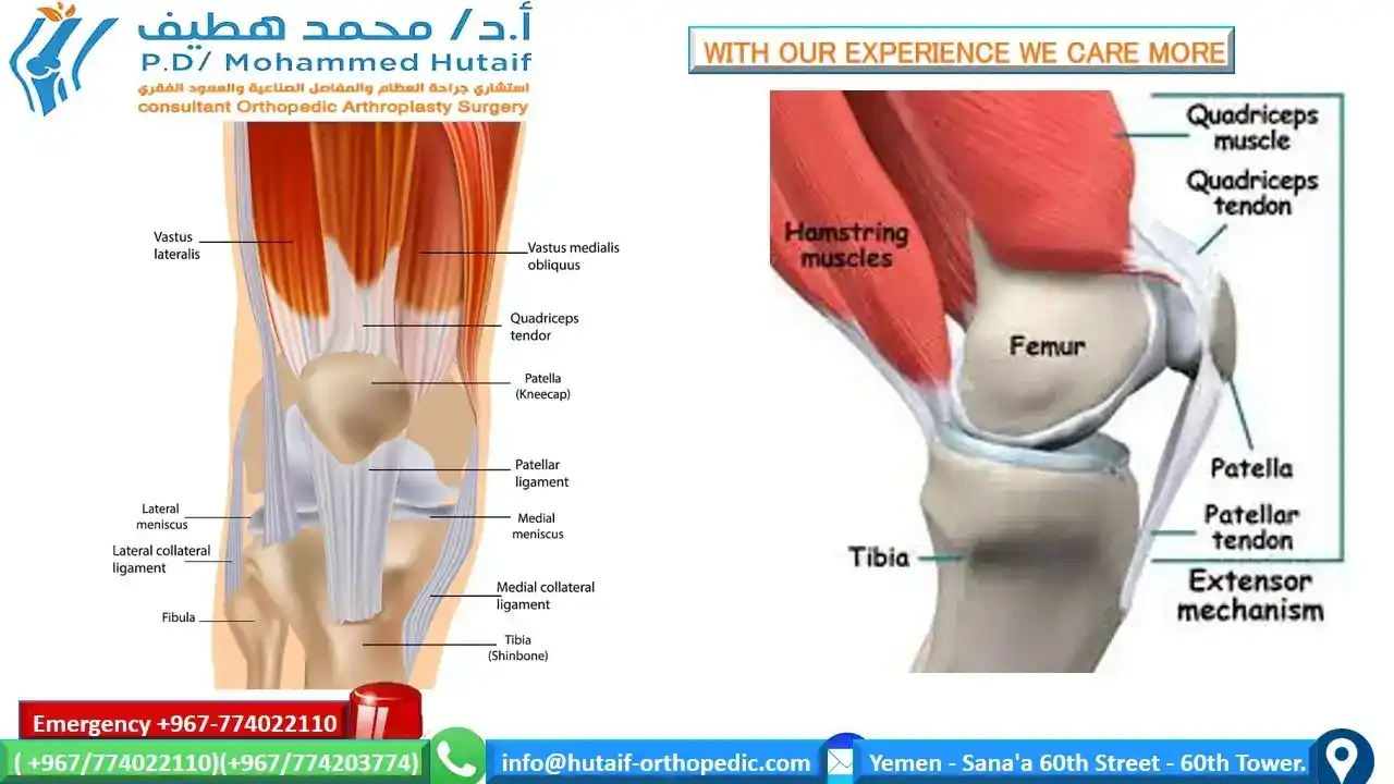
For the knee, this is fairly simple. We need a relatively short list of parts: 4 bones, 2 tendons, 4 ligaments, and 2 types of cartilage.
To start, we place the femur, tibia, and fibula bones in their proper positions.
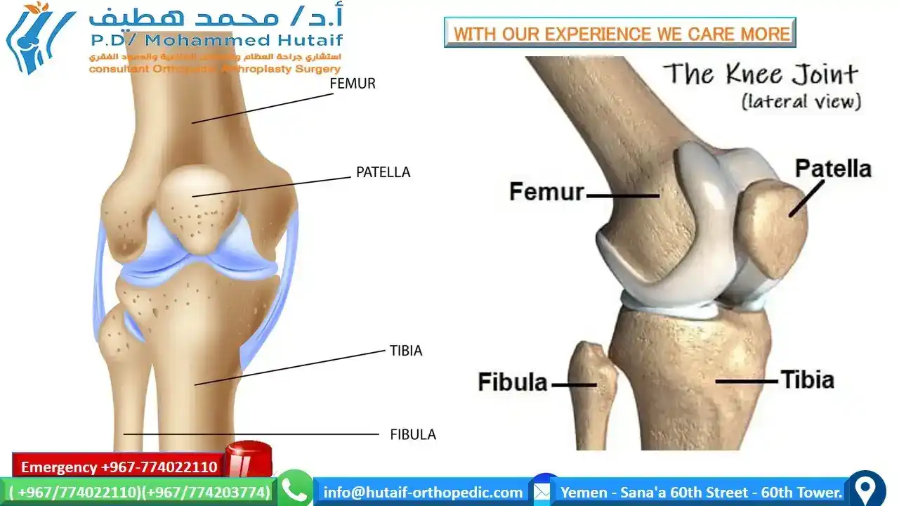
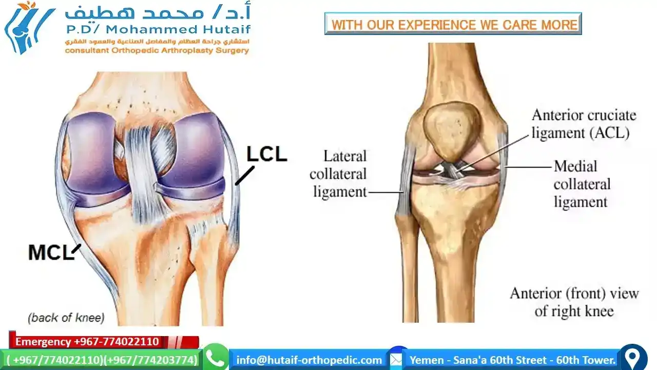
Next, we need a system of ligaments to hold them together) and a coating of articular cartilage on the surface of the femur and tibia, two of the three bones that will articulate against each other
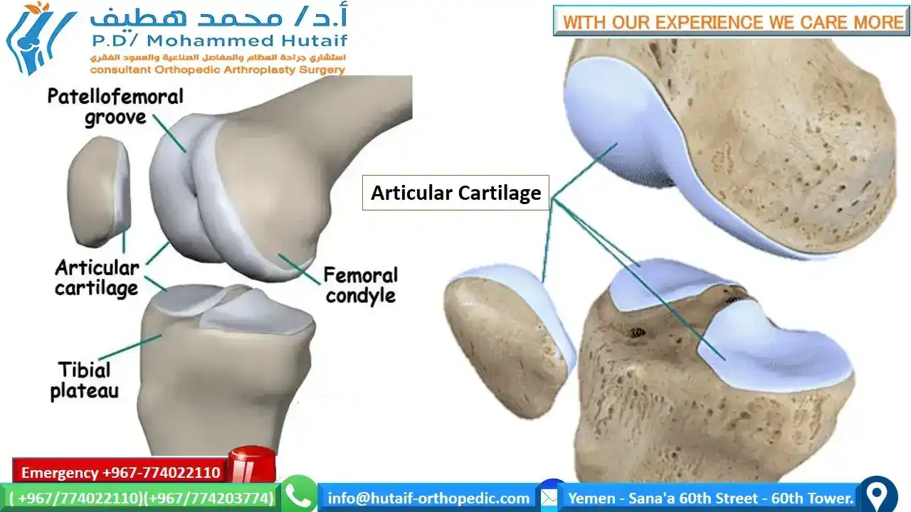
Of all the structures used to assemble the knee we are building, this thin layer of glistening articular cartilage tissue is probably the most important and the most interesting
Now we are ready to add the meniscus cartilages, which sit like two rubbery, horseshoe-shaped pads on the surface of the tibia The exact role that the meniscus cartilages play in the function of the knee is poorly understood, but the two wedge-shaped pieces of fibrocartilage act as shock absorbers between your femur and tibia. These are the menisci. The menisci help to transmit weight from one bone to another and play an important role in knee stability
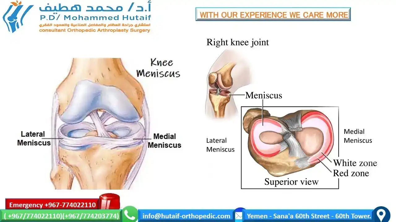
The menisci increase stability for femorotibial articulation, distribute axial load, absorb shock, and provide lubrication and nutrition to the knee joint
The last bone we need to add if we are building a knee is the patella. The patella is a link in the chain of structures known as the extensor mechanism. These structures—the quadriceps muscle, the quadriceps tendon, the patella, and the patellar tendon—allow us to forcibly straighten (extend) our knees. When it contracts, the quadriceps muscle (via its quadriceps tendon attachment to the patella) pulls the patella proximally. As it is pulled proximally, the patella (via its patella tendon attachment to the tibia) pulls the anterior tibia proximally, which rotates the knee into extension.
