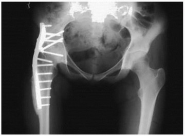
Hip Arthrodesis
INTRODUCTION
The average age of patients presenting with symptomatic coxarthrosis is decreasing (1). Excellent medium- to long-term results can be predictably achieved in such patients with total hip arthroplasty (THA). The results of THA in young, active patients (less than 50 years old) are less predictable. Failure rates in this group have been reported as higher (2,3), and risks particular to arthroplasty such as dislocation, osteolysis, and soft tissue reactions to wear particles (adverse reaction to metal debris) are genuine concerns. These results are expected to improve with newer techniques and technology.
The surgeon thus needs to consider all available options in this patient population, which would allow return to an active, productive, and independent lifestyle while delaying their need for a THA. Nonsurgical treatment in this group often fails to address the cause of their symptoms and can sentence them to an increasingly dependent and sedentary lifestyle with resultant health and socioeconomic consequences.
INDICATIONS FOR HIP ARTHRODESIS
The ideal candidate is a teenage or young adult male patient involved, or likely to be, in heavy manual labor.
CONTRAINDICATIONS
Contraindications to arthrodesis include the following:
- Active infection
- Lower back pain or evidence of an abnormality that may become painful (e.g., spondylolisthesis)
- Ipsilateral knee instability or osteoarthritis
- Contralateral hip osteoarthritis
- Significant bone loss (absent head and neck) or poor quality such that rigid fixation would be challenging

PREOPERATIVE ASSESSMENT AND SURGICAL PLANNING
Appropriate patient selection is vital. The most common causes for conversion of arthrodesis to THA include progressive pain in the lumbar spine, ipsilateral knee, and contralateral hip (9). As a result, concurrent degenerative disease in these areas needs to be excluded with a thorough history examination and radiographic evaluation. It is important to accurately measure and document leg lengths and to explain that modest shortening of the limb is to be expected after operation. This is in fact desirable because it facilitates swing through of the foot while walking.

POSTOPERATIVE CARE
Feather weight-bearing mobilization is started on day 1 postsurgery. If a drain is used, this is removed 24 to 48 hours post-op.
Partial weight bearing commences at 6 weeks and full weight bearing at 3 months post surgery. Patients are usually discharged at 3 to 5 days. They are placed on an oral anticoagulant for 35 days.
Radiologic signs of bone union are usually seen at 3 to 4 months. A visible joint space, radiolucent lines around the screws, and pain at 6 months or more suggest a nonunion.

COMPLICATIONS
Complications occur in 15% to 50% of patients having hip arthrodesis. These include malposition, nonunion, leg length discrepancy, failure to relieve pain, and patient dissatisfaction.

