
Managing Mid-shaft Clavicle Fracture: A Comprehensive Guide

Mid-shaft Clavicle Fracture
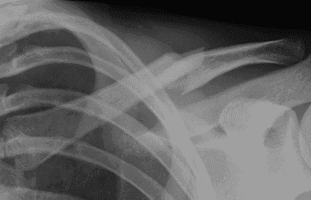
The X-ray demonstrates a comminuted junction of mid and distal third fracture with significant displacement and overlap of the fracture ends. However, the Coracoclavicular distance, acromioclavicular joint, and glenohumeral joint appear normal. This indicates a complex fracture with multiple fragments, which may require surgical management for optimal outcome.
Click here to see more
Clavicle Fractures - Epidemiology and Anatomy
EPIDEMIOLOGY
Clavicle fractures account for approximately 2.6% of all fractures and for 44% to 66% of fractures about the shoulder. Middle third fractures account for 80% of all clavicle fractures, whereas fractures of the lateral and medial third of the clavicle account for 15% and 5%, respectively.
ANATOMY
The clavicle is the first bone to ossify (fifth week of gestation) and the last ossification center (sternal end) to fuse, at 22 to 25 years of age. The clavicle is S-shaped, with the medial end convex forward and the lateral end concave forward. It is widest at its medial end and thins laterally.
The medial and lateral ends have flat expanses that are linked by a tubular middle, which has sparse medullary bone. The clavicle functions as a strut, bracing the shoulder from the trunk and allowing the shoulder to function at optimal strength.
The medial one-third protects the brachial plexus, the subclavian and axillary vessels, and the superior lung. It is strongest in axial load. The junction between the two cross-sectional configurations occurs in the middle third and constitutes a vulnerable area to fracture, especially with axial loading. Moreover, the middle third lacks reinforcement by muscles or ligaments distal to the subclavius insertion, resulting in additional vulnerability. The distal clavicle contains the coracoclavicular ligaments. The two components are the trapezoid and conoid ligaments. They provide vertical stability to the acromioclavicular (AC) joint. They are stronger than the AC ligaments.
CLASSIFICATION
| Classification | Description | ||||||||||||
|---|---|---|---|---|---|---|---|---|---|---|---|---|---|
| Allman |
Group I: fracture of the middle third (80%). This is the most common fracture in both children and adults; proximal and distal segments are secured by ligamentous and muscular attachments. 
|
||||||||||||
|
Group II: fracture of the distal third (15%). This is subclassified according to the location of the coracoclavicular ligaments relative to the fracture:

|
|||||||||||||
|
Group III: fracture of the proximal third (5%). Minimal displacement results if the costoclavicular ligaments remain intact. It may represent epiphyseal injury in children and teenagers. Subgroups include:
|
|||||||||||||
| ORTHOPAEDIC TRAUMA ASSOCIATION |
Classification of clavicle fractures. See Fracture and Dislocation Classification Compendium at: https://ota.org/research/fracture-and-dislocation-compendium |
TREATMENT
NONOPERATIVE
Most minimally displaced clavicle fractures can be successfully treated nonoperatively with some form of immobilization. Comfort and pain relief are the main goals. A sling has been shown to provide the same results as a figure-of-eight bandage, providing more comfort and fewer skin problems.
The goals of the various methods of immobilization are as follows:
- To support the shoulder girdle, raising the lateral fragment in an upward, outward, and backward direction (sling)
- To depress the medial fragment (figure of eight)
- To maintain some degree of fracture reduction (both)
- To allow for the patient to use the ipsilateral hand and elbow
Regardless of the method of immobilization utilized, some degree of shortening and deformity usually result. In general, immobilization is used for 4 to 6 weeks. During the period of immobilization, active range of motion of the elbow, wrist, and hand should be performed.
OPERATIVE
The surgical indications for midshaft clavicle fractures have become more standard in the past 20 years. The accepted indications for operative treatment of acute clavicle fractures are open fracture, associated neurovascular compromise, and skin tenting with the potential for progression to open fracture.
Controversy exists over management of midshaft clavicle fractures with substantial displacement (more than 100%), comminution (Z deformity), and shortening (>1 to 2 cm). Although most displaced midshaft fractures will unite, studies have reported shoulder dysfunction and patient dissatisfaction with the resulting cosmetic deformity. There is also more recent evidence that functional outcome may be improved in some of these patients with surgical treatment. Furthermore, the presence of a malunion may portend inferior function.
Controversy also exists over management of type II distal clavicle fractures. Some authors have indicated that all type II fractures require operative management due to high rate of nonunion. Others report that if the bone ends are in contact, healing can be expected even if there is some degree of displacement. In this situation, nonoperative management consists of sling immobilization and progressive range of shoulder motion.
Operative fixation may be accomplished via the use of:
- Plate fixation: This is placed either on the superior or on the anteroinferior aspect of the clavicle. Plate and screw fixation requires a more extensive exposure than intramedullary devices but has the advantage of more secure fixation counteracting tensile forces. Plate and screw fixation may be prominent, particularly if placed on the superior aspect of the clavicle. Adjunctive suture fixation (substitution) of the coracoclavicular ligaments may be helpful for distal fractures with limited area for screw purchase. Newer low-profile implants and/or anteroinferior placement may preclude this finding.
- Intramedullary fixation: usually placed in retrograde fashion through the lateral fragment and then in anterograde fashion into the medial fragment or antegrade as a flexible implant that is then stiffened. Use of intramedullary fixation requires frequent radiographic follow-up to monitor the possibility of hardware migration and a second procedure for hardware removal. Older intramedullary pins are prone to skin erosion at the hardware insertion site laterally. Historically, these implants have been reported to be associated with complications in up to 50% of cases.
- Operative treatment of type II distal clavicle fractures consists of reducing the medial fragment to the lateral fragment. This is accomplished by using either coracoclavicular fixation (Mersilene tape, sutures, wires, or screws) or fixation across the AC joint, through the lateral fragment and into the medial fragment (lateral clavicle plates).
COMPLICATIONS
Neurovascular compromise: This is uncommon and can result from either the initial injury or secondary to compression of adjacent structures by callus and/or residual deformity. Subclavian vessels are at risk with superior plating.
Malunion: This may cause a bony prominence, and resultant shortening may be associated with poorer Disabilities of the Arm, Shoulder and Hand (DASH) scores at 1 year.
Nonunion: The incidence of nonunion following clavicle fractures ranges from 0.1% to 13.0%, with 85% of all nonunions occurring in the middle third. Factors that have been implicated in the development of nonunions of the clavicle include: severity of initial trauma (open wound), extent of initial displacement of fracture fragments, soft tissue interposition, refracture, inadequate period of immobilization, primary open reduction and internal fixation.
Posttraumatic arthritis: This may occur after intra-articular injuries to the sternoclavicular or AC joint.
Click here to see more
Mid-shaft Clavicle Fracture
The X-ray demonstrates a comminuted junction of mid and distal third fracture with significant displacement and overlap of the fracture ends. However, the Coracoclavicular distance, acromioclavicular joint, and glenohumeral joint appear normal. This indicates a complex fracture with multiple fragments, which may require surgical management for optimal outcome.