Pediatric Diaphyseal Fractures of the Tibia and Fibula
Pediatric diaphyseal fractures of the tibia and fibula - Epidemiology, Anatomy, Mechanism of Injury, Clinical Evaluation, Radiographic Evaluation, Treatment, Complications
Epidemiology
Of pediatric tibial fractures, 39% occur in the middle third.
Approximately 30% of pediatric diaphyseal fractures are associated with a fracture of the fibula. Occasionally, this is in the form of plastic deformation, producing valgus alignment of the tibia.
Isolated fractures of the fibular shaft are rare and result from direct trauma to the lateral aspect of the leg.
Anatomy
The nutrient artery arises from the posterior tibial artery, entering the posterolateral cortex distal to the origination of the soleus muscle, at the oblique line of the tibia. Once the vessel enters the intramedullary canal, it gives off three ascending branches and one descending branch. These give rise to the endosteal vascular tree, which anastomoses with periosteal vessels arising from the anterior tibial artery.
The anterior tibial artery is particularly vulnerable to injury as it passes through a hiatus in the interosseous membrane.
The peroneal artery has an anterior communicating branch to the dorsalis pedis artery.
The fibula is responsible for 6% to 17% of weight-bearing load. The common peroneal nerve courses around the neck of the fibula, which is nearly subcutaneous in this region; it is therefore especially vulnerable to direct blows or traction injuries at this level.
Mechanism of Injury
- Direct: Trauma to the leg occurs, mostly in the form of vehicular trauma or pedestrian–motor vehicle accident.
- Indirect: In younger children, most tibial fractures result from torsional forces. These spiral and oblique fractures occur as the body mass rotates on a planted foot. The fibula prevents significant shortening when intact, but the fracture frequently falls into varus.
Clinical Evaluation
The patient typically presents with pain, swelling, and tenderness in the region of the fracture.
Motion of the knee is painful, and the child usually refuses to ambulate.
Children with stress fractures of the tibia may complain of pain on weight bearing that is partially relieved by rest.
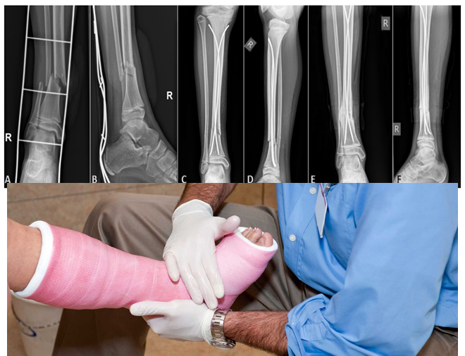
Radiographic Evaluation
Standard AP and lateral views of the leg should be obtained.
Radiographs of the ipsilateral ankle and knee should be obtained to rule out associated injuries.
Comparison views of the uninjured, contralateral leg may be obtained in cases in which the diagnosis is unclear.
MRI may be obtained to rule out occult fracture.
Classification
Descriptive
- Angulation
- Displacement
- Open versus closed
- Pattern: transverse, oblique, spiral, greenstick, plastic deformation, torus
- Comminution
Treatment
Nonoperative
Most pediatric fractures of the tibia and fibula are uncomplicated and may be treated by simple manipulation and casting, especially when they are nondisplaced or minimally displaced. However, isolated tibial diaphyseal fractures tend to fall into varus, whereas fractures of the tibia and fibula tend to fall into valgus with shortening and recurvatum.
Displaced fractures may be initially treated with closed reduction and casting with the patient under general anesthesia.
In children, acceptable reduction includes 50% apposition of the fracture ends, <1 cm of shortening, and <5- to 10-degree angulation in the sagittal and coronal planes with <5 degrees of rotation.
A long leg cast is applied with the ankle slightly plantar flexed (20 degrees for distal and middle third fractures, 10 degrees for proximal third fractures) to prevent posterior angulation of the fracture in the initial 2 to 3 weeks. The knee is flexed to 45 degrees to provide rotational control and to prevent weight bearing.
Fracture alignment must be carefully monitored, particularly during the initial 3 weeks. Atrophy and diminished swelling may result in loss of reduction. Some patients require repeat manipulation and cast application under general anesthesia 2 to 3 weeks after initial casting.
The cast may require wedging (opening or closing wedge) to provide correction of angulatory deformity. If the anticipated wedge is to be greater than 15 degrees, it is advisable to change the cast.
Time to healing varies according to patient age:
- Neonates: 2 to 3 weeks
- Children: 4 to 6 weeks
- Adolescents: 8 to 12 weeks
Operative
Operative management of tibial fractures in children are typically required in <5% of cases.
Indications for operative management include:
- Open fracture
- Fractures in which a stable reduction is unable to be achieved or maintained
- Associated vascular injury
- Fractures associated with compartment syndrome
- Severely comminuted fractures
- Associated femoral fracture (floating knee)
- Fractures in patients with spasticity syndromes (cerebral palsy, head injury)
- Patients with bleeding diatheses (hemophilia)
- Patients with multisystem injuries
Open fractures or grossly contaminated fractures with associated vascular compromise may be treated with debridement of compromised tissues and external fixation, particularly in older children. Regional or free flaps or skin grafting may be required for skin closure.
Other methods of operative fixation include percutaneous pins, plates and screws, flexible or “elastic” intramedullary nails or rigid intramedullary nails (in adolescents, after closure of the proximal tibia physis).
Postoperatively, a long leg cast is usually placed (depending on the method of fixation), with the knee in 15 degrees of flexion to allow for rotational control. The cast is maintained for 4 to 16 weeks depending on the status of healing, as evidenced by serial radiographs, as well as the healing of associated injuries.
Complications
Compartment syndrome: In pediatric tibia fractures, compartment syndrome is most common after severe injury in which the interosseous membrane surrounding the anterior compartment is disrupted. Patients with elevated compartment pressures >30 mm Hg or within 30 mm Hg of diastolic blood pressure should receive emergency fasciotomies of all four compartments of the leg to avoid neurologic and ischemic sequelae.
Angular deformity: Correction of deformity varies by age and gender.
- Girls <8 years old and boys <10 years old often experience significant remodeling.
- Girls 9 to 12 years old and boys 11 to 12 years old can correct up to 50% of angulation.
- In adolescents >13 years, <25% angular correction is expected.
- Posterior and valgus angulation tends to correct the least with remodeling.
Malrotation: Rotational deformity of the tibia does not correct with remodeling and is poorly tolerated, often resulting in malpositioning of the foot with the development of associated ankle and foot problems. Supramalleolar osteotomy may be required for rotational correction.
Premature proximal tibial physeal closure: This may occur with unrecognized crush injury (Salter–Harris type V) to the proximal tibial physis, resulting in growth arrest. This most commonly affects the anterior physis and leads to a recurvatum deformity of the affected knee.
Delayed union and nonunion: Uncommon in children, but it may occur as a result of infection, the use of external fixation, or inadequate immobilization. Fibulectomy, bone grafting, reamed intramedullary nailing (adolescents), and plate fixation with bone grafting have all been described as methods to treat tibial nonunions in the pediatric population.
1. What is the most common mechanism of injury for pediatric tibial fractures?
Answer: b) Indirect torsional forces. In younger children, most tibial fractures result from torsional forces. These spiral and oblique fractures occur as the body mass rotates on a planted foot.
2. What is the nutrient artery's entry point into the tibia?
Answer: d) Posterior tibial artery, at oblique line of the tibia. The nutrient artery arises from the posterior tibial artery, entering the posterolateral cortex distal to the origination of the soleus muscle, at the oblique line of the tibia.
3. What is the treatment for severely comminuted pediatric tibial fractures?
Answer: b) External fixation. Operative management of tibial fractures in children are typically required in <5% of cases. Indications for operative management include severely comminuted fractures, open fractures, fractures associated with compartment syndrome, and more. Grossly contaminated fractures with associated vascular compromise may be treated with debridement of compromised tissues and external fixation, particularly in older children.
4. What should be monitored during the initial 3 weeks of casting for pediatric tibial fractures?
Answer: d) Atrophy and loss of reduction. Fracture alignment must be carefully monitored, particularly during the initial 3 weeks. Atrophy and diminished swelling may result in loss of reduction. Some patients require repeat manipulation and cast application under general anesthesia 2 to 3 weeks after initial casting.
5. What may occur with unrecognized crush injury to the proximal tibial physis in pediatric tibial fractures?
Answer: d) Premature physeal closure. Premature proximal tibial physeal closure may occur with unrecognized crush injury (Salter–Harris type V) to the proximal tibial physis, resulting in growth arrest. This most commonly affects the anterior physis and leads to a recurvatum deformity of the affected knee.