Understanding The Anatomy of The vertebral Column: A Comprehensive Guide
Learn about the structure and function of the vertebrae, spinal nerves, and back muscles while gaining insights into how the body supports itself and how to maintain an appropriate posture.
Table of Contents:
- I. Introduction
- II. Extrinsic and Intrinsic Back Muscles
- III. Structure of the Vertical Column
- IV. Naming Convention
- V. Anatomical Landmarks
- VI. Curvatures
- VII. Specialized Joints
- VIII. Conclusion
I. Introduction
The vertical column is part of the axial skeleton and functions to support the body's weight and encloses and protects the spinal cord within the spinal canal. It provides a framework for the attachment of several muscles.
II. Extrinsic and Intrinsic Back Muscles
The extrinsic back muscles are involved in moving the upper limbs and ribcage, whereas the intrinsic back muscles are important in maintaining posture and in moving the vertical column itself.
III. Structure of the Vertical Column
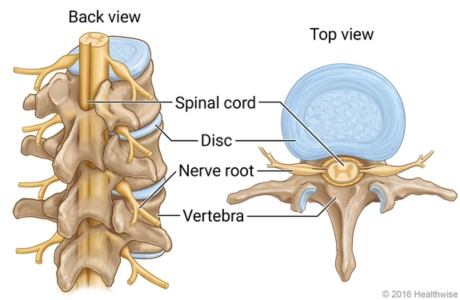
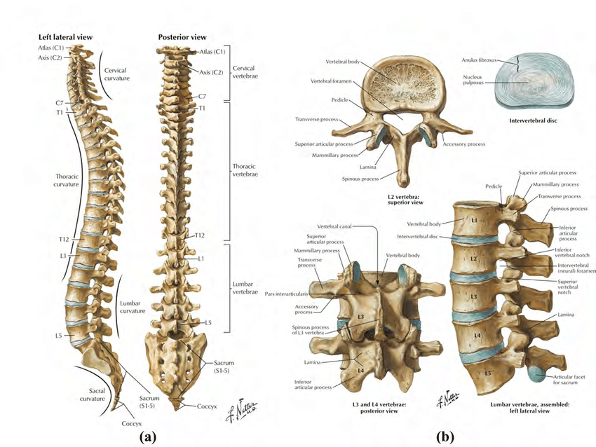
The vertical column consists of 24 articulating vertebrae and then the sacrum and coccyx which consist of nine fused vertebrae. In between each vertebrae there is an intervertebral disc which forms a fibrocartilaginous joint allowing slight movement between the vertebrae and functioning as a shock absorber for the spine. The intervertebral disc consists of a tough outer fibrous ring called the annulus fibrosis and a gelatinous center, the nucleus pulposus.
There are seven cervical vertebrae, 12th thoracic, 5 lumbar, 5 sacral, and three to four coccygeal.
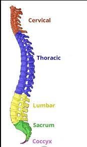
IV. Naming Convention
The vertebrae are identified by a letter and a number. For example, for the cervical vertebrae you have vertebrae C1 to C7, for the thoracic vertebrae you have T1 to T12, and for the lumbar vertebrae you have L1 to L5. This naming convention is useful when referring to anatomical landmarks and when referring to spinal nerves.
With regard to the spinal nerves, these are named according to the level at which they exit the vertical column within the cervical spine. Although there are only seven vertebrae, there are actually eight spinal nerves.
V. Anatomical Landmarks
In terms of using spinal levels for reference points to anatomical landmarks, a clinical example would be in the context of performing a lumbar puncture. In adults, we know that the spinal cord terminates at the conus medullaris which lies at approximately the L1 to L2 level. We can palpate the iliac crests to top of which we now lie at approximately the L4 level. From here, we can identify the spinous process of L4 and a safe entry point for the needle to avoid damage to the spinal cord.
Another example would include the sternal angle of Louis or the manubriosternal junction which lies at the T4/T5 level. This level determines the thoracic plane, an artificial horizontal plane which divides the superior mediastinum from the inferior mediastinum and at which there are a few key anatomical landmarks such as the start and end of the aortic arch and bifurcation of the trachea.
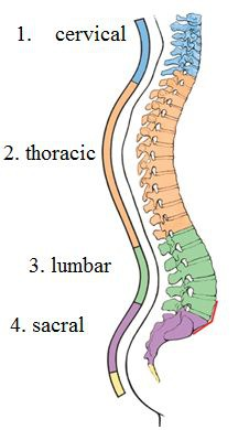
VI. Curvatures
As you've seen, the adult vertical column is curved with four curvatures: the cervical lordosis which is convex anteriorly, the thoracic kyphosis which is convex dorsally, the lumbar lordosis which is convex anteriorly, and the pelvic curvature which is concave anteroposteriorly starting at the lumbosacral junction and ending at the coccyx.
In neonates, the vertical column just has one fixed kyphotic curvature which is concave anteriorly and known as the primary curvature. The other lordotic curvatures are progressively developed later in life as shown in this diagram and are known as secondary curvatures.
VII. Specialized Joints
There are two more specialized joints within the cervical spine: the atlanto-occipital joint and the atlanto-axial joint. The atlanto-occipital joint is given this name because the first vertebra is also referred to as the atlas and the second vertebra is known as the axis. This joint enables flexion and extension and a small amount of lateral flexion. The atlanto-axial joint, on the other hand, is a complex joint which consists of a specialized synovial pivot joint formed between the odontoid peg of C2 and the anterior arch of C1. This unique anatomical configuration enables rotational movement.
VIII. Conclusion
Understanding the anatomy of the vertical column is important for healthcare professionals and anyone interested in human anatomy. By learning about the structure and function of the vertebrae, spinal nerves, and back muscles, you can gain a better appreciation for how the body supports itself and how to maintain proper posture. Thank you for reading!
The Intrinsic and Extrinsic Back Muscles: Everything You Need To Know
The intrinsic and extrinsic back muscles play a crucial role in maintaining posture and movement. Learn about their functions and the importance of maintaining a healthy spine.