A Comprehensive Guide to the Muscles of the Leg
In this guide, we'll break down the muscles of the leg by compartment, including the posterior, anterior, and lateral compartments. We'll also discuss innervation and function of each muscle.
So the leg is the region of the lower limb, which lies between the knee joint and the ankle joint.
So I'm going to break this tutorial into two parts, so I'm going to do the first part on the posterior compartment, and then the second part will be on the muscles of the anterior and lateral compartments.
So just like my tutorials on the thigh and the upper limb, the muscles of the leg can be broken down into compartments.
And these compartments are separated by intermuscular septa, and the interosseus membrane between the tibia and the fibula.
Posterior Compartment
The muscles of the posterior compartment here, mainly act to plantar flex the foot, flex the digits, and invert the foot.
So plantar flexion is when you, get up on your tip toes basically, so if you, so if you can see this angle between the foot and the shin, it...you extend this angle.
So you open this angle further, so you, so it's getting up on your tip toes essentially, and dorsiflexion is bringing your toes towards your head.
So if you imagine lying down on your back and bringing your toes up towards your head - that's dorsiflexion and plantarflexion is the opposite.
The muscles of the posterior compartment plantar flex at the ankle joint, they flex the digits, and they invert the foot.
The muscles of the anterior compartment, here, do the opposite really, so they dorsiflex the foot, so bring the toes upwards.
They extend the digits, and they invert the foot also.
So as well as plantarflexion, flexion of the digits and inversion, there are two muscles in the posterior compartment which actually can flex at the knee because of their attachment on the femur, and these are the gastrocnemius and the plantaris muscle, which I'll come on to talk about.
The lateral compartment are the muscles here, which lie laterally, and these evert the foot.
This tutorial will be concerned with the muscles of the posterior compartment of the leg.
This article focuses on the muscles in the posterior compartment of the leg, including the gastrocnemius, soleus, and popliteus muscles. We'll dive into their origin, insertion, and primary functions, including the ability to flex the knee.
So briefly just a quick word about innervation.
If I just bring in the nerves, you can see, so just looking here at the popliteal fossa, you can see the sciatic nerve, and it splits into two main branches, so you've got the tibial branch of the sciatic nerve, and you've got the common peroneal branch, the common fibular branch of the sciatic nerve.
So the tibial branch of the sciatic nerve supplies muscles of the posterior compartment, and the common peroneal nerve which winds round laterally here, innervates the anterior and lateral compartments of the leg.
So just a quick point about the common fibular nerve, just while I've mentioned it.
So if I just remove the muscle layer, you can see the relationship of this nerve with the head of the fibula, so this nerve winds round the...well the neck of the fibula, and this, at this point, it's very vulnerable so any impact or fractures can easily damage this nerve, and because this nerve supplies the anterior and lateral compartments of the leg, it results in foot drop.
So it's worth making a note of that point, so that's the common fibula nerve, which winds round laterally around the neck of the fibula and it's quite exposed in this region.
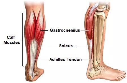
Muscles of the Posterior Compartment
The muscles of the posterior compartment can be separated into superficial and deep muscles, and in total you've got seven muscles.
So you've got three muscles in the superficial layer, and four muscles in the deep layer.
So I'll just work from superficial to deep and talk you through these structures.
So obviously we're looking at the most superficial muscle here, and this is called the gastrocnemius muscle.
So this muscle has two heads, it's got a medial head here, and a lateral head.
And this muscle inserts onto the femur on the medial and lateral condyles.
So the medial head inserts on the medial condyle and the lateral head inserts on the lateral condyle.
So this muscle has two functions, it plantarflexes the foot, so plantarflexion is when you get up on your tip toes, so you can see how this would act if you look at the insertion, so you can see if the muscle contracts it pulls the calcaneus upwards and it would get you up on your tip toes - so that's plantarflexion.
And also because of its origin on the femoral condyles, it can also flex at the knee.
So that's the gastrocnemius muscle and that's the most superficial muscle.
So next we've got this tiny little muscle here, which is called the plantaris muscle,
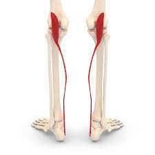
So this muscle actually lies under the medial head of the gastrocnemius muscle.
So you can see the tendon emerging here and it actually lies underneath this.
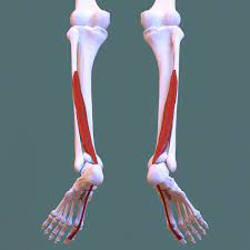
So next we've got this large muscle here called the soleus muscle.
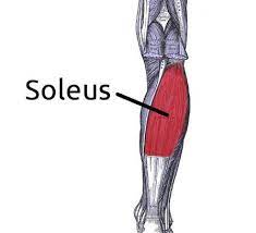
So this muscle doesn't originate on the femur, so it can't flex the knee, so this muscle's primary function is to plantar flex at the ankle joint.
So these three muscles are innervated by the tibial nerve - so that's the branch of the sciatic nerve which innervates the posterior compartment.
So next we have the muscles of the deep layer,
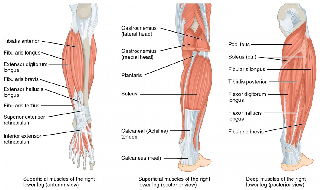
The most superior muscle is the popliteus muscle,
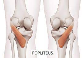
So this muscle actually serves to unlock the knee when it's locked in extension.
So it does this by laterally rotating the femur, so when it contracts, so its origin here, insertion up here - so when it contracts, it brings the femur round, laterally rotating it and unlocking the knee.
So in the standing position, the knee is fully extended and it's locked like this.
So next we've got these three large muscles.
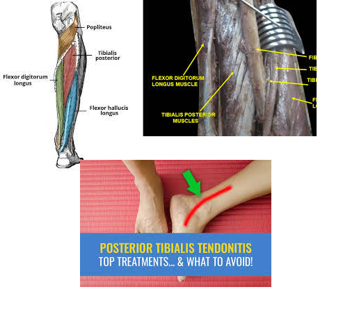
Starting with the most lateral muscle - the flexor hallucis longus,
So next we've got this muscle here which is called the flexor digitorum longus,
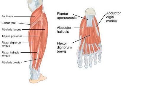
So the last muscle is this muscle which sits between them - the tibialis posterior muscle.
So those are the muscles of the posterior compartment of the leg, I hope that's cleared things up a little bit.
Do you know the difference between the superficial and deep muscles of the leg? In this article, we'll explore each layer and its corresponding muscles, including the tibialis posterior and the flexor digitorum longus.