Ankle Fractures in Children: Classification, Treatment, and Prognosis
Learn about the classification, treatment options, and prognosis of ankle fractures in children. Understand the importance of proper management and how it can impact long-term outcomes
Ankle Fractures in Children
Ankle fractures in children make up about 5% of all fractures in this age group and are the second most common type of fracture after distal radius fractures. They account for approximately 15% of all physeal fractures. Nonoperative treatment is usually the preferred method for managing these fractures, but in certain cases surgical treatment may be necessary. Surgical treatment is typically recommended when there is significant misalignment of the joint or angular deformity.
Classifying pediatric ankle fractures can be helpful in determining the appropriate treatment and predicting the outcome. The Salter-Harris method is the most commonly used classification system for physeal fractures. The Lauge-Hansen classification, which is typically used for adult ankle fractures, can also be modified for pediatric fractures. Studies have shown that pronation-type injuries are more likely to result in premature closure of the growth plate compared to supination-external rotation (SER) type injuries. This classification system is widely known among orthopedic surgeons. Other classification systems include the Danis-Weber system and a more comprehensive classification proposed by Dias and Tachdjian, which combines the Lauge-Hansen and Salter-Harris classifications.
Transitional fractures of the ankle, which occur during skeletal maturity, are caused by the uneven closure of the distal tibial growth plate. Triplane fractures are complex fractures that consist of sagittal, transverse, and coronal components with an epiphyseal and metaphyseal fragment. Tillaux fractures are most common in adolescents within the first year after the closure of the distal tibial growth plate. They occur when an external rotational force causes the anterolateral aspect of the tibial epiphysis, which is attached to the anterior inferior tibiofibular ligament, to be avulsed. This ligament is stronger than the remaining open growth plate.
The ligaments of the ankle provide stability to the ankle joint. They play a role in the development of transitional fractures (triplane and Tillaux fractures) because they are often stronger than the growth plate, making children more prone to physeal fractures rather than ankle sprains.
The anteroinferior tibiofibular ligament is attached to the anterolateral border of the tibial epiphysis. When an external rotation force is applied to the foot, it can cause the avulsion of the anterolateral fragment of the tibial epiphysis. The strength imbalance between the ligament and the weaker growth plate can lead to transitional Tillaux and triplane fractures.
Understanding the anatomy of the distal tibial growth plate is important for managing and predicting the outcome of ankle fractures. The closure of the growth plate usually occurs around 15 years of age in girls and 17 years of age in boys, and understanding the specifics of physeal closure can help determine appropriate intervention and management.
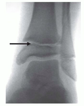
FIG 1 • Bump in the center. The arrow shows the Kump bump, which is where the closure of the growth plate begins. We speculate that damage to this structure can cause the growth plate to close prematurely.
The perichondral ring of LaCroix is an area between the articular cartilage and the periosteum of the diaphysis. It is made up of perichondrium and has the potential to produce cartilage and bone. The perichondral ring provides stability to the growth plate and may play a role in certain fractures and growth plate injuries in children. The adjacent periosteum, which is firmly attached to the perichondral ring, can become trapped in the fracture site and hinder proper alignment.
The Lauge-Hansen classification system was created in 1950 to understand how fractures occur by replicating fracture patterns in cadavers. This system grades the severity of ankle injuries as I to IV for rotational patterns and I to II for translational patterns.
A recent study of pediatric ankle fractures found that SER fractures accounted for 66%, abduction fractures accounted for 30%, pronation-external rotation fractures accounted for 3%, and axial crush injuries accounted for 1%. The causes of these ankle fractures varied, with most occurring due to falls and sports activities. Abduction injuries during soccer or skateboarding were found to have a higher likelihood of developing premature physeal closure compared to other activities.
Certain fractures in adolescence are influenced by the specific anatomy and patterns of growth plate closure. For instance, the same external rotation mechanism can lead to either a Tillaux fracture or a triplane fracture, depending on the age and degree of physeal closure in the child. The lateral part of the distal tibial physis typically closes last, making it a common site for weakness in skeletally maturing children. This weakness allows for an anterolateral fragment to be avulsed from the epiphysis, resulting in Tillaux fractures or the intraarticular fragment in triplane fractures.
The occurrence of premature physeal closure in distal tibial fractures has been historically considered rare, with reported incidences as low as 2% to 5%. However, recent data reveals an overall incidence of physeal arrest in Salter-Harris type I and II fractures to be 38%, and as high as 55% for displaced fractures. The mechanism of injury and the treatment approach have been shown to influence the rate of premature closure. SER injuries have a lower overall incidence of premature physeal closure (38%) compared to abduction-type injuries (52%).
These findings have important implications for the prognosis of pediatric ankle fractures. Previous research has demonstrated a 3.5-fold increase in the premature physeal closure rate for Salter-Harris types I and II fractures if a gap of 3 mm or more is present on postreduction imaging. Presence of periosteum in physis can lead to residual fracture gapping and premature physeal closure. Physicians should discuss the possibility of premature physeal closure with the patient's family during the initial visit, particularly in cases of abduction-type injuries.
The initial evaluation of a child's ankle injury involves understanding the mechanism and timing of the injury. The examination should include an assessment of the skin and soft tissues, identification of areas of tenderness upon palpation, and a thorough sensory, motor, and vascular examination. In the diagnosis of ankle fractures in children, it is important to consider conditions such as osteomyelitis and child abuse.
The presence of periosteum in the physis results in residual fracture gapping and contributes to premature physeal closure. Orthopaedic surgeons should discuss the possibility of premature physeal closure with the patient's family during the initial visit, particularly in cases of abduction-type injuries.
If an ankle injury is suspected, a complete radiographic ankle series should be obtained, including anteroposterior (AP), lateral, and mortise views. The mortise view is particularly important for identifying anterolateral fragments in Tillaux fractures, which may not be well visualized on the AP or lateral view due to overlap from the posterior fibula.
We do not recommend stress radiographs for children, though an external rotation stress view may be used intraoperatively to evaluate for syndesmosis injury in children nearing skeletal maturity. Contralateral comparison films can be useful in differentiating accessory ossicles from fractures. Computed tomography (CT) is often necessary to fully understand many ankle fractures, and we often recommend three-dimensional postreduction CT scans. CT scans are especially important for assessing residual displacement on plain radiographs after attempted closed reduction.
Possible alternative diagnoses for ankle injuries include ankle sprain, accessory ossicles, osteochondritis dissecans (osteochondral lesions), contusions, and osteomyelitis.
Nonoperative treatment is usually the preferred method for managing these fractures, but in certain cases surgical treatment may be necessary. Our approach to surgical management recommends attempting closed reduction of closed ankle fractures under conscious sedation in the emergency department. Reductions are typically performed using ketamine for conscious sedation and with the assistance of a portable image intensifier. The reduction technique should reverse the mechanism of injury determined from the patient's history and fracture pattern, such as the Quigley maneuver for abduction-external rotation fractures. After reduction, patients are placed in fiberglass casts.
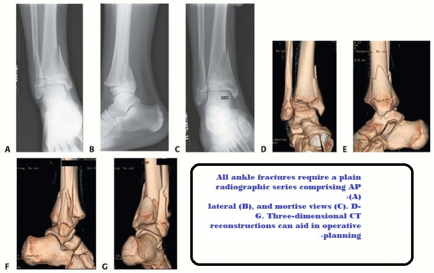
FIG 3 • Short-leg fiberglass cast after reduction of distal tibial physeal fracture is univalved, spacers are later placed, and the cast is overwrapped with waterproof tape.
For Salter-Harris types I and II fractures and triplane fractures, if near-anatomic reduction with less than 2 mm of residual displacement is achieved, we proceed with a long-leg cast and non-weight bearing for 4 weeks. Radiographs are periodically taken during this time, with frequency determined by the stability of the fracture pattern. For cases with significant residual displacement, we opt for open reduction and internal fixation.
Key Points on Ankle Fractures in Children
- Ankle fractures in children make up about 5% of all fractures in this age group and are the second most common type of fracture after distal radius fractures.
- Surgical treatment is typically recommended when there is significant misalignment of the joint or angular deformity.
- The Salter-Harris method is the most commonly used classification system for physeal fractures.
- Pronation-type injuries are more likely to result in premature closure of the growth plate compared to supination-external rotation (SER) type injuries.
- The ligaments of the ankle provide stability to the ankle joint and play a role in the development of transitional fractures.
- The anatomy of the distal tibial growth plate is important for managing and predicting the outcome of ankle fractures.
- The Lauge-Hansen classification system helps understand how fractures occur by replicating fracture patterns in cadavers.
- The occurrence of premature physeal closure in distal tibial fractures has been historically considered rare, but recent data reveals a higher incidence.
- The mechanism of injury and the treatment approach influence the rate of premature closure.
- The initial evaluation of a child's ankle injury involves understanding the mechanism and timing of the injury and assessing the skin and soft tissues.
- An external rotation stress view may be used intraoperatively to evaluate for syndesmosis injury in children nearing skeletal maturity.
- Complete radiographic ankle series should be obtained, including anteroposterior (AP), lateral, and mortise views.
- Contralateral comparison films can be useful in differentiating accessory ossicles from fractures.
- CT scans are often necessary to fully understand many ankle fractures.
- Other possible alternative diagnoses for ankle injuries include ankle sprain, accessory ossicles, osteochondral lesions, contusions, and osteomyelitis.
- Nonoperative management involves attempting closed reduction, placing patients in fiberglass casts, and monitoring for complications.
- If near-anatomic reduction is achieved, long-leg cast and non-weight bearing are recommended.
- Open reduction and internal fixation may be required for cases with residual intra-articular irregularity or significant angular deformity.