Myositis Ossificans: A Guide to Understanding and Treating this Bone-Forming Condition
Introduction
Myositis Ossificans is a reactive soft tissue bone-forming process that commonly occurs following a traumatic event to soft tissues. It is characterized by the formation of peripheral bone within the soft tissues, often causing pain, tenderness, swelling, and decreased range of motion. This condition typically affects patients between the ages of 15 and 35, with males being more commonly affected. In this comprehensive guide, we will delve deeper into the key aspects of Myositis Ossificans, including its epidemiology, etiology, clinical presentation, diagnostic imaging, and treatment options.
Epidemiology
Myositis Ossificans is most commonly observed in young active males between the ages of 15 and 35. The condition tends to affect certain anatomic locations more frequently, such as the quadriceps, brachialis, and gluteal muscles. The condition is less common in females and older age groups.
Etiology
Myositis Ossificans is a form of heterotopic ossification, which refers to the formation of bone in abnormal locations. It is often the result of direct trauma to the soft tissues or an intramuscular hematoma. The most common location for the development of Myositis Ossificans is the diaphysis of long bones. It is essential to differentiate Myositis Ossificans from tumors, as fibrodysplasia ossificans progressiva (FOP), a rare subtype of heterotopic ossification, involves a mutation of the ACVR1 gene.
Genetics
Myositis Ossificans is almost always a posttraumatic condition, meaning it occurs following an injury. Genetic factors do not play a significant role in its development.
Presentation
Patients with Myositis Ossificans typically experience pain, tenderness, swelling, and decreased range of motion in the affected area. These symptoms usually manifest within days of the traumatic event. Over time, the pain and size of the mass decrease, but the mass itself can continue to grow for several months, reaching a size of 3 to 6 cm. Once the growth phase ceases, the mass becomes firm. Physical examination reveals a palpable soft tissue mass and restricted range of motion.
Imaging

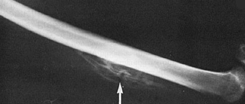
Radiographs are commonly used for the diagnosis of Myositis Ossificans. They show peripheral bone formation with a central lucent area, often resembling a "dotted veil" pattern. MRI with gadolinium can also be employed, where rim enhancement is visualized within the first three weeks. CT scan imaging reveals an eggshell appearance of the lesion.
Histology
Characteristic histology of Myositis Ossificans presents a zonal pattern. The periphery of the lesion shows mature trabeculae of lamellar and woven bone, which can be visualized through calcification on X-ray. In contrast, the center of the lesion consists of an irregular mass of immature fibroblasts, potentially accompanied by a cartilage component. No cellular atypia is typically seen.
Treatment
Nonoperative treatment options for Myositis Ossificans include rest, range of motion exercises, and activity modification. However, it is important to note that passive stretching is contraindicated, as it may exacerbate the condition. Physical therapy can be utilized to maintain range of motion, while radiographic monitoring confirms the maturation of the lesion. In cases where the condition remains problematic even after maturation, surgical excision is indicated. However, operating during the acute phase is not recommended, and it is advisable to wait at least six months before considering excision. It is worth mentioning that excising the lesion within 6 to 12 months predisposes to local recurrence.
Prognosis
Myositis Ossificans is a self-limiting condition, meaning it typically resolves on its own with time. The mass usually begins to decrease in size after one year, indicating a favorable prognosis for patients with this condition.
Conclusion
Myositis Ossificans is a reactive soft tissue bone-forming process that occurs following trauma to soft tissues. It predominantly affects young active males and is commonly observed in the quadriceps, brachialis, and gluteal muscles. Diagnosis is made through radiographs showing peripheral bone formation with central lucent areas, while treatment options include nonoperative approaches such as rest, range of motion exercises, and physical therapy. Surgical excision is reserved for persistently symptomatic lesions. With its favorable prognosis and self-limiting nature, Myositis Ossificans is a condition that can be effectively managed with proper care and monitoring.

