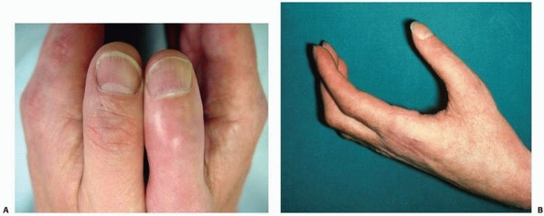
Late Phase of Complex Regional Pain Syndrome
which affects every tissue. The skin is thinned and joint creases and
subcutaneous fat disappear. Hairs become fragile, uneven, and curled,
while nails are pitted, ridged, brittle, and discolored brown. Palmar
and plantar fascias thicken and contract simulating Dupuytren disease.106
Tendon sheaths become constricted, causing triggering and increased
resistance to movement. Muscle contracture combined with tendon
adherence leads to reduced tendon excursion. Joint capsules and
collateral ligaments become shortened, thickened, and adherent, causing
joint contracture.
is very variable. Within orthopaedic practice, the large majority of
patients who demonstrate the features of the early phase of CRPS after
trauma will not go on to develop severe late phase contracture,
although a significant proportion will show chronic subclinical
contracture.106
Bone involvement is universal with increased uptake on bone scanning in early CRPS (Fig. 23-3). This was originally thought to be periarticular, suggesting arthralgia84,97,110; however, CRPS does not cause arthritis and more recent studies have shown generalized hyperfixation,5,34,40 confirming the view of Doury et al.44 Increased uptake is not invariable in children.167
Later, the bone scan returns to normal and there are radiographic
features of rapid bone loss: visible demineralization with patchy,
subchondral or subperiosteal osteoporosis, metaphyseal banding, and profound bone loss98 (Fig. 23-4). Despite the osteoporosis, fracture is uncommon, presumably because the patients protect the painful limb very effectively.
are extremely rare. Thus, severe, chronic CRPS associated with severe
contracture is uncommon with a reported prevalence of less than 2% in
retrospective series.8,75,102,108,129
In contrast, prospective studies designed to look specifically for the
early features of CRPS show that they occur after 30% to 40% of every
fracture and surgical trauma (e.g., total knee replacement),2,3,7,13,14,51,81,135,139 where the features of CRPS have been actively sought. Furthermore, statistically, the features tend to occur together.3 These common early cases of CRPS are usually not specifically diagnosed.139 They resolve substantially either spontaneously or with standard treatment by physical therapy and analgesia within 1 year.13,14,105,139
Some features, particularly stiffness, may remain, suggesting that CRPS
may be responsible for significant long-term morbidity even when mild.5,18 The truly intriguing question is, if CRPS is so common, why is it not a universal finding after trauma or orthopaedic surgery?
identical stimulus in a different limb does not cause it. The incidence
is not changed by treatment method and open anatomic reduction and
rigid internal fixation does not abolish it.135 It is unclear whether injury severity or quality of fracture reduction alters the incidence.3,14 There is, however, an association with excessively tight casts55 and there may be a genetic predilection.41,94,96,111,112 The following etiologies have been proposed:
Most orthopaedic clinicians immediately recognize a “Sudecky”
patient—that is, broadly speaking, a patient who appears to the
clinician to be somebody who is likely to fare poorly after surgical
intervention or trauma, perhaps because of to their inability to
cooperate fully with physical therapy. In fact, the literature fails to
identify this sort of patient and the evidence does not support the
notion that CRPS is primarily psychological.25 Studies of premorbid personality show no consistent abnormality.122,172 Most patients are psychologically normal,158 although emotional lability, low pain threshold,39 hysteria,127 and depression144 have been reported. There is an association with antecedent psychological stress,20,25,63,64,65,156 which probably exacerbates pain in CRPS, as in other diseases.23
It seems likely that the severe chronic pain of CRPS causes depression
and that a “Sudecky” type of patient who develops CRPS is at risk of a
poor outcome because they will not mobilize in the face of pain.
CRPS is characterized by excessive and abnormal pain. Pain is usually caused when an intense noxious stimulus activates high
threshold nociceptors, thus preventing tissue damage. Neuropathic pain
in CRPS occurs without appropriate stimulus and has no protective
function. However, injured peripheral nerve fibers undergo cellular
changes, which cause usually innocuous tactile inputs to stimulate the
dorsal horn cells via A-β fibers from low-threshold mechanoreceptors,
causing allodynia in CRPS 2.92,167
Similar C-nociceptor dysfunction explains causalgia. Furthermore,
axonal injury prevents nerve growth factor transport, which is
essential for normal nerve function.104,168 In CRPS 1, covert nerve lesions with artificial synapses have been postulated.43
These “ephases” have not been demonstrated and are unnecessary since
inflammatory mediators released by the initial trauma (and possibly
retained due to a failure of free radical clearance), can sensitize
nociceptors to respond to normally innocuous stimuli.168
include abnormalities in skin blood flow, temperature regulation and
sweating, and edema. However, SNS activity is not usually painful.88,89 In CRPS, however, some pain (termed sympathetically maintained pain [SMP]141)
is SNS dependent. This accounts for spontaneous pain and allodynia,
which may therefore be relieved by stellate ganglion blockade130 and then restored by noradrenalin injection.1,148
Furthermore, there is an abnormal difference in cutaneous sensory
threshold between the limbs, which is reversed by sympathetic blockade,54,57,131,132 while increasing sympathetic activity worsens pain.90
the body’s reaction to injury. After partial nerve division, injured
and uninjured somatic axons express α-adrenergic receptors30 and sympathetic axons come to surround sensory neuron cell bodies in dorsal root ganglia.117,161,168 These changes, which may be temporary,148,159,160
make the somatic sensory nervous system sensitive to circulating
catecholamines and norepinephrine released from postganglionic
sympathetic terminals.
leading to gross scarring. For this reason, the major differential
diagnoses within an orthopaedic context are occult causes of
inflammation such as soft tissue infection or stress fracture. Indeed,
CRPS is associated with inflammatory changes including macromolecule
extravasation125 and reduced oxygen consumption.71,149 In animals, infusion of free radical donors causes a CRPS-like state,150 and amputated human specimens with CRPS show basementmembrane thickening consistent with overexposure to free radicals.151 These considerations suggest that CRPS is an exaggerated local inflammatory response to injury.72,73
In other words, on this hypothesis, CRPS represents a local form of the
systemic free radical disease that causes adult respiratory distress
syndrome and multiple organ failure after severe trauma. This concept
is supported by evidence that the free radical scavenger vitamin C is
effective prophylaxis against post-traumatic CRPS.170,171
changes in early CRPS is a primary capillary imbalance causing stasis,
extravasation, and consequent local tissue anoxia.48,49,114,134
It is a common clinical observation that patients who appear to be at
risk of developing CRPS are unable or unwilling to cooperate with
physical therapy to mobilize their limb after trauma or orthopaedic
surgery. Indeed, undue immobilization has traditionally been believed
to be at least an important contributory factor in the generation of
CRPS or even the sole cause.9,47,121,163
afferent sensory perception but only recently has the possibility of
abnormal efferent motor function been systematically explored.
Classically, it was believed that the “immobile RSD limb” was guarded
by the patient to prevent inadvertent painful movement or sensory
contact.44,60
In fact, CRPS is associated with an abnormality of motor function that
is often overlooked partially because of patient embarrassment and
partly because in the past it has been labeled as “hysterical.”33,152 In 1990, Schwartzman and Kerrigan137
reported a subgroup of CRPS patients with a variety of motor disorders
and a minority of patients with CRPS demonstrate obvious dystonia or
spasms.10,45,110,113
A prospective study of 829 CRPS patients showed that abnormalities of
motor function were reported by 95%, varying from weakness to
incoordination and tremor.30
Objective testing in small numbers of patients shows that CRPS patients
have impaired grip force coordination, target reaching, and grasping.136,164
reasons for the lack of movement in CRPS. Patients demonstrate evidence
of “neglect” of the affected limb, similar to that seen after parietal
lobe stroke. When asked about moving the limb, statements are made such
as “my limb feels disconnected from my body” and “I need to focus all
my mental attention and look at the limb in order for it to move the
way I want… .”59 Another study
revealed bizarre perceptions about a body part including a desperate
desire for amputation. There was a mismatch between limb sensation and
appearance with mental erasure of the affected part. These authors
suggested the term “body perception disturbance” rather than “neglect”
to describe this phenomenon. 101 There appears to be a central sensory
confusion, in that when a nonnoxious stimulus is provided that the
patient finds painful due to allodynia, the patient is unable to
determine whether it is truly painful, and by impairing integration
between sensory input and motor output, movement is impaired.83,115
limb and find it difficult to initiate or accurately direct movement
and there is a mismatch between sensation, perception, and movement.29,60,152
Failure to use the limb appears to relate to this rather than the
traditional view of learned pain avoidance behavior in response to
allodynia. Whatever the exact cause, failure of mobilization may be
central to the etiology of CRPS because all the features of phase 1
CRPS, except pain, are produced in volunteers after a period of cast
immobilization.27,28,29 This may be explained by the fact that activity-dependent gene function is common in the nervous system.168 and normal tactile and proprioreceptive input are necessary for correct central nerve signal processing.103
The rationale for MVF is restoration of the congruence between sensory
and motor information, and it was originally used for the treatment of
phantom limb pain.133 The patients
are instructed to exercise both the unaffected and the affected limb.
However, a mirror is placed so that they cannot see the affected limb,
and when they think they are looking at it, they are actually observing
the mirror image of their normal limb. As might be expected, MVF
resulted in improvement in range of movement; however, in addition in
early CRPS, MVF also abolished or substantially improved pain and
vasomotor instability.150


