
Hip Dysplasia-Description-PATHOGENESIS
Developmental Dislocation (Dysplasia) of the Hip (DDH)-Description-PATHOGENESIS
■In all cases of DDH, the socket (acetabulum) is shallow, meaning that the ball of the thighbone (femur) cannot firmly fit into the socket.
■Sometimes, the ligaments that help to hold the joint in place are stretched.
■The degree of hip looseness, or instability, varies among children with DDH.
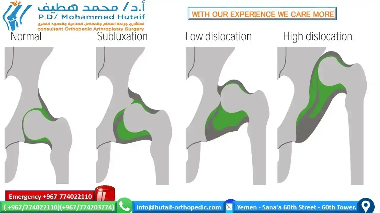
Dislocated. In the most severe cases of DDH, the head of the femur is completely out of the socket.
Dislocatable. In these cases, the head of the femur lies within the acetabulum, but it can easily be pushed out of the socket during a physical examination.
Subluxatable. In mild cases of DDH, the head of the femur is simply loose in the socket. During a physical examination, the bone can be moved within the socket, but it will not dislocate.
Illustrations of a normal hip and a dislocated hip
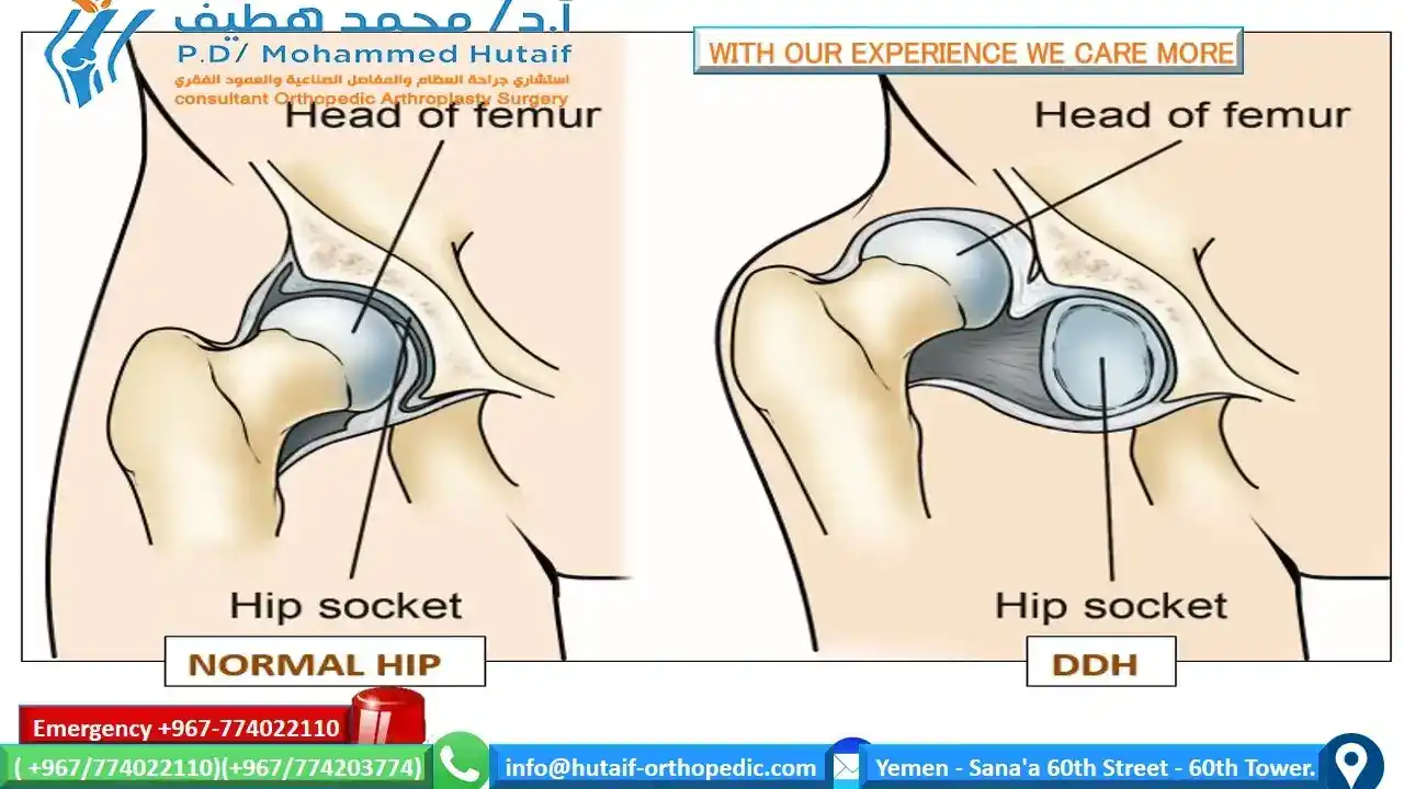
(Left) In a normal hip, the head of the femur fits firmly inside the hip socket. (Right) In severe cases of DDH, the thighbone is completely out of the hip socket (dislocated).
■In the United States, approximately 1 to 2 babies per 1,000 are born with DDH.
■Pediatricians screen for DDH at a newborn's first examination and at every well-baby checkup thereafter.
PATHOGENESIS
■ Because of an abnormality in the growth centers of the acetabulum, abnormal periosteal growth, or abnormal positioning of the femoral head, the acetabulum does not develop
properly.
■ Even with a concentric reduction of the femoral head, the
prior period of abnormal growth may prevent the acetabulum
from achieving a normal configuration at maturity. The older
the child is at the time of reduction, the more likely an osteotomy will be necessary.
■DDH has also been called congenital dislocation (and dysplasia) of the hip (CDH). both names correctly describe some components of the condition, and they often use the names interchangeably.
■“Congenital dysplasia of the hip” implies that the hip is abnormal at birth, distinguishing this condition from other diseases that cause dysplasia and dislocation in childhood, such as cerebral palsy, polio, muscular dystrophy, and other neuromuscular diseases.
■“Developmental dysplasia of the hip” emphasizes that the etiology is a developmental abnormality that results in a hip disorder with a wide spectrum of problems, ranging from instability to complete (frank) dislocation.
■DDH is currently the most popular name for this condition